44 onion cells under microscope with labels
Onion Epidermal Cell Labeled Diagram - schematron.org Draw a labelled diagram of an onion epidermal cell seen under the microscope. ( 4 marks) e The onion epidermal cells are not green in colour because they lack. The epidermal cells of onions provide a protective layer against viruses and fungi that may harm the sensitive tissues. PDF Onion Cell Lab - SomeWaresInMaine 1. Onion layer (tissue) 2. iodine stain 3. slide & cover slip Procedure: 1. Carefully separate the thin film tissue from between two layers of an onion 2. Carefully place a small sample of this tissue onto a slide - avoid folds & creases 3. Put a drop of iodine stain on the tissue 4. Carefully place a coverslip to avoid air bubbles 5.
Blog, She Wrote - Embracing the Independent & Authentic ... Blog, She Wrote - Embracing the Independent & Authentic ...
Onion cells under microscope with labels
Animal Cell Diagram Under Microscope Labeled Animal Cell Diagram Under Microscope Labeled. Animal Cell Diagram Under Microscope. Function cell does in the body dictate the change and adaptation done by cell. When observing onion cells, there is the Cell Surface Membrane which is present in all living cells. We all keep in mind that the human body is quite intricate and a method I ... Observing Onion Cells Under The Microscope Afterwards, carefully mount the prepared and stained onion cell slide onto the microscope stage. Make sure that the cover slip is perfectly aligned with the microscope slide, and that any excess stain has been wiped off. Secure the slide on the stage using the stage clips. Labeled Onion Cell Under Microscope 40x - Micropedia Labeled onion cell under microscope 40x. While photosynthesis takes place in the leaves of an onion containing chloroplast the little glucose that is produced from this process is converted in to starch starch granules and stored in the bulb. This slide was scanned using a 40x 080na objective. Machine vt recommended for you.
Onion cells under microscope with labels. Onion cells under the microscope: 40X - 100X - 400X - YouTube under the #microscope: 40X - 100X - 400X Onion Root Tip Mitosis - Stages, Experiment and Results · Cover the sample (root tip) with a coverslip and gently press the coverslip down, then examine the slide under the microscope starting with low magnification * For this experiment, a properly prepared slide should appear light pink due to the stain to almost colorless. * Unused roots can be stored in 70 percent alcohol. Results Onion Cells Under a Microscope - Requirements/Preparation ... Add a drop of iodine solution on the onion membrane (or methylene blue) Gently lay a microscopic cover slip on the membrane and press it down gently using a needle to remove air bubbles. Touch a blotting paper on one side of the slide to drain excess iodine/water solution, Place the slide on the microscope stage under low power to observe. Microscope Cell Lab: Cheek, Onion, Zebrina - SchoolWorkHelper The onion epidermis cell is the only cell that has a cell wall. In addition, it is the only cell that has a chloroplast, where photosynthesis can happen. The cheek epithelium cell is the only one that has centrioles, the barrel-shaped organelle that is responsible for helping organize chromosomes during cell division.
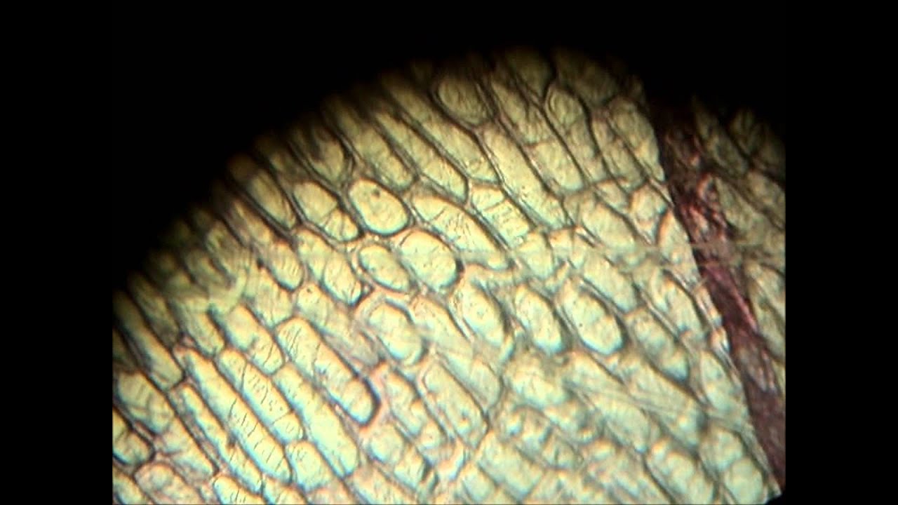
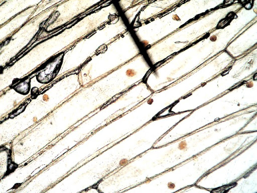
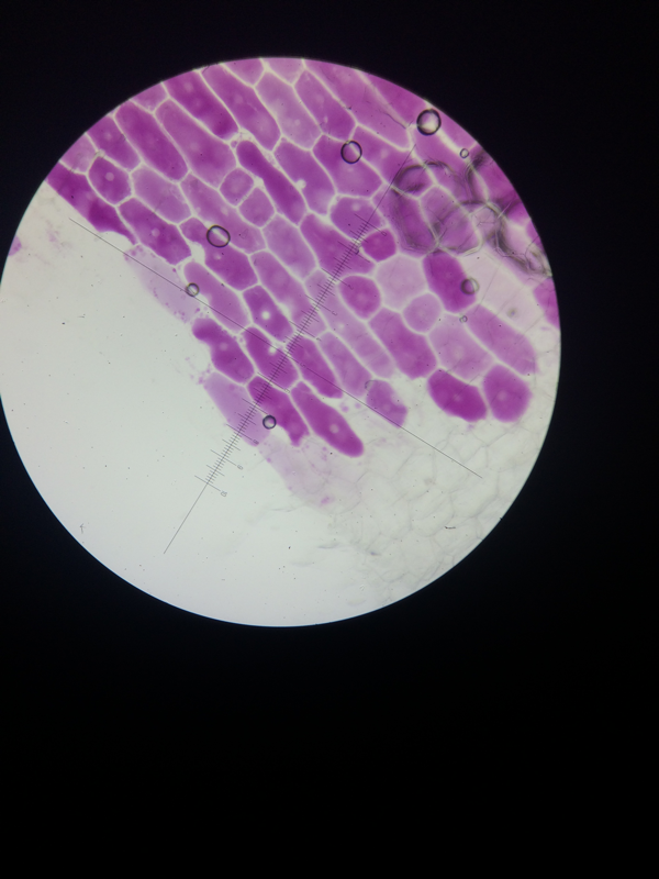
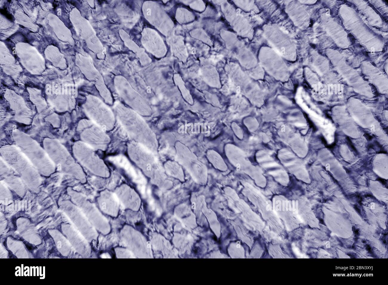
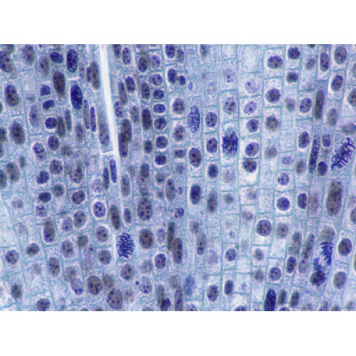
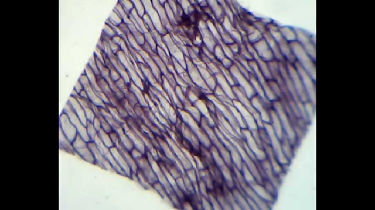
Onion Skin Epidermis Sample under microscope 4x ... - YouTube A sample on an onion skin epidermis diyed in blue for visibility, viewd under the microscope at 4x and 10x magnification.microscope:Biolux model :AL Animal Cell Under Light Microscope Labelled : Draw and ... Onion cell diagram labeled structure of animal cell and plant cell under microscope. An organelle found in large numbers in most cells, in which the biochemical processes of respiration and energy production occur. Under a light microscope, the cell membrane, nucleus and cytoplasm of a cheek cell (animal cell) can be observed. Onion Root Mitosis - Microscopy-UK It is common to see photomicrographs of onion root cells when demonstrating how cell division takes place in plants. Onions have larger chromosomes than most plants and stain dark. The chromosomes are easily observed through a compound light microscope. The cells pictured below are located in the apical meristem of the onion root. The apical ... Onion Cell Lab Report.docx - Onion Cell Lab Report By Onion Cell Lab Report By : Nawaf Almalki Introduction: Many things that are viewed using a microscope, particularly cells, can appear quite transparent under the microscope. The internal parts of the cells, the organelles, are so transparent that they are often difficult to see. Biologists have developed a number of stains that help them see the cells and their organelles by adding color to ...
animal cell under microscope labeled - Rayford Runyon When observing onion cells there is the Cell Surface Membrane which is present in all living cells. Most cells both animal and plant range in size between 1 and 100 micrometers and are thus visible only with the aid of a microscope. While observing with tissues or on tissue. Labeled Onion Cell Under Microscope 40x - Micropedia Labeled onion cell under microscope 40x. While photosynthesis takes place in the leaves of an onion containing chloroplast the little glucose that is produced from this process is converted in to starch starch granules and stored in the bulb. This slide was scanned using a 40x 080na objective. Machine vt recommended for you. Observing Onion Cells Under The Microscope Afterwards, carefully mount the prepared and stained onion cell slide onto the microscope stage. Make sure that the cover slip is perfectly aligned with the microscope slide, and that any excess stain has been wiped off. Secure the slide on the stage using the stage clips. Animal Cell Diagram Under Microscope Labeled Animal Cell Diagram Under Microscope Labeled. Animal Cell Diagram Under Microscope. Function cell does in the body dictate the change and adaptation done by cell. When observing onion cells, there is the Cell Surface Membrane which is present in all living cells. We all keep in mind that the human body is quite intricate and a method I ...
Onion Cells Under Microscope! REALLY COOL!!! - YouTube

Cells Zone – I'm a Scientist, Get me out of Here!

How to Observe Onion Cells under a Microscope | Things under a microscope, Science cells, High ...
Biology Pictures: Onion Cells under Microscope
Light Microscope Onion Cell Labeled - Micropedia
Onion Cells High Resolution Stock Photography and Images - Alamy

Cell structure | Cells as the basic units of life | Siyavula
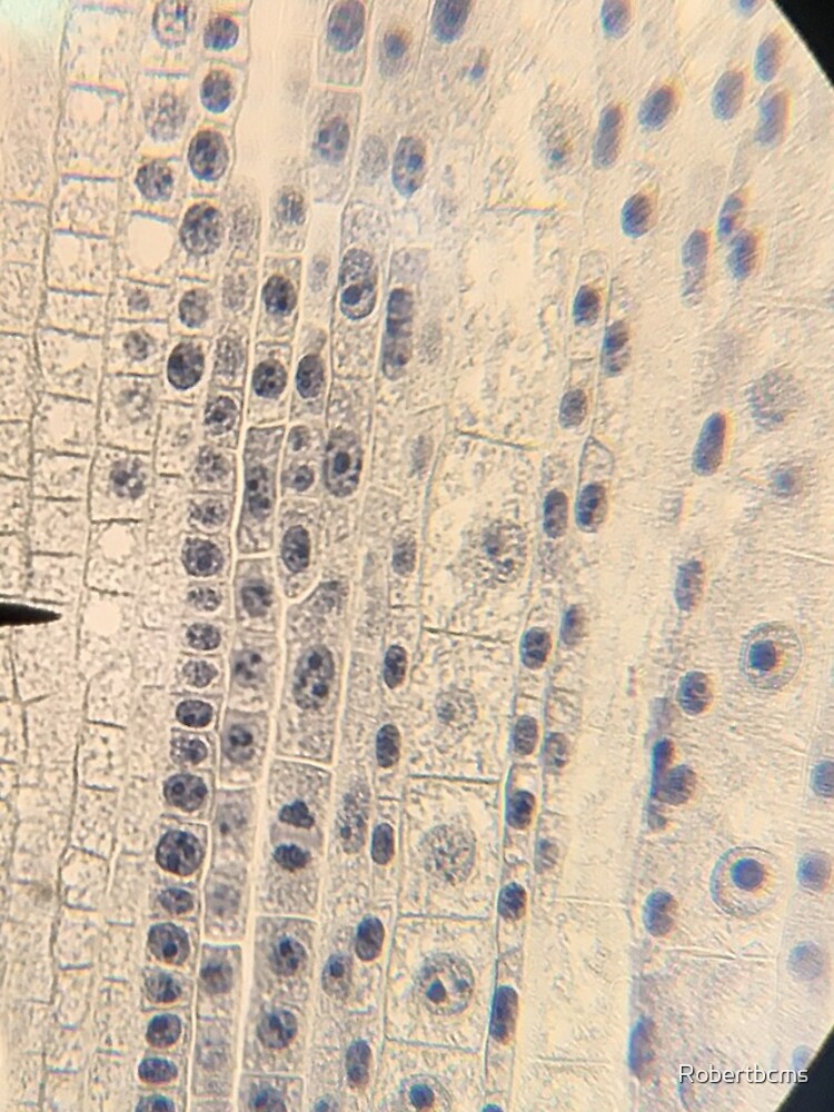
"Biology- onion root tip cells under microscope" iPhone Case & Cover by Robertbcms | Redbubble
Slide, Microscope, Onion Root Tip Mitosis

Rens blog : Science, cells

swifty science: onion cell lab

Red Onion Cell Under Microscope Labeled - Micropedia

Biology 1 - Onion cell microscopy - YouTube
Onion cells under the microscope: 40X - 100X - 400X - YouTube
Cells: Microscope cheek and onion cell | Teaching Resources

Fanos' MCB Blog: Onion Skin
Post a Comment for "44 onion cells under microscope with labels"