43 the brain with labels
Related 3D Models for 3d model the brain with labels ID: 700274 , 1,984 views , Tags: 3d models, anatomy, brain, head, human, label, medical Description: This is a 3d model of the brain with labels,which tells us the information of the brain.This model is sliced into parts frontal lobe, motor cortex, etc. Illustration Picture of Brain Anatomy - Brain - eMedicineHealth Picture of Brain. The brain is the complex organ responsible for processing sensory information (sound, touch, taste, sight, and smell). The brain controls voluntary and involuntary movements. Signals from the brain tell muscles to contract. Input from the brain controls the function of other organs in the body.
Brain Label | Human anatomy and physiology, Basic anatomy and ... Brain Nervous System This picture of the neuron is unlabeled, write in the labels to test your knowledge of the anatomy of a neuron. L Mary Lanier School Science Education Behavioral Science Health Education Physical Education Philosophy Of Mind Cognitive Psychology Structural features of a typical nerve cell (i.e., neuron) and synapse.

The brain with labels
Labeled Diagrams of the Human Brain You'll Want to Copy Now Labeled Diagrams of the Human Brain Central Core The central core consists of the thalamus, pons, cerebellum, reticular formation and medulla. These five regions are the central areas that regulate breathing, pulse, arousal, balance, sleep and early stages of processing sensory information. 3D Brain This interactive brain model is powered by the Wellcome Trust and developed by Matt Wimsatt and Jack Simpson; reviewed by John Morrison, Patrick Hof, and Edward Lein. Structure descriptions were written by Levi Gadye and Alexis Wnuk and Jane Roskams . Label The Brain Worksheets & Teaching Resources | TpT The Primary Brain. 89. $2.50. Digital Download. ZIP (19.68 MB) These table caddy labels measure 8" by 3" in size and are completely EDITABLE! They can be used as classroom labels for a table caddy, table numbers, name tags, or anything else you need this size label for.
The brain with labels. The Human Brain - Visible Body Rotate this 3D model to see the four major regions of the brain: the cerebrum, diencephalon, cerebellum, and brainstem. The brain directs our body's internal functions. It also integrates sensory impulses and information to form perceptions, thoughts, and memories. The brain gives us self-awareness and the ability to speak and move in the world. the brain with labels brain labels human inside labeled Lateral View Of The Brain Centered At The Level Of The Intraparietal brain sulcus lateral intraparietal neuroanatomy centered level What Are The 4 Main Types Of Electrical Injury? - Pat Labels electrical types Parts of the brain: Learn with diagrams and quizzes | Kenhub Labeled brain diagram First up, have a look at the labeled brain structures on the image below. Try to memorize the name and location of each structure, then proceed to test yourself with the blank brain diagram provided below. Labeled diagram showing the main parts of the brain Blank brain diagram (free download!) Brain Label (Remote) - The Biology Corner The activity includes an external view of the brain where students label the lobes of the cerebrum (frontal, parietal, occipital, and temporal) and the cerebellum. Next students drag and drop labels to the internal structures, such as the thalamus, midbrain, corpus callosum, pineal body, and colliculi.
Brain (Human Anatomy): Picture, Function, Parts, Conditions, and More The brain is also divided into several lobes: • The frontal lobes are responsible for problem solving and judgment and motor function. • The parietal lobes manage sensation, handwriting, and body... Labels on the Brain - Cognitioneducation We love labels for what they do well — they make things easy for us. Labels are a product of the way our minds work - in fact, this process may be one of our brains greatest feats. Our brain circuitry pattern-matches like nobody's business; doing so affords a necessary level of simplicity in an otherwise overly complex world. Brain Anatomy and How the Brain Works - Hopkins Medicine The cerebellum ("little brain") is a fist-sized portion of the brain located at the back of the head, below the temporal and occipital lobes and above the brainstem. Like the cerebral cortex, it has two hemispheres. The outer portion contains neurons, and the inner area communicates with the cerebral cortex. Label The Brain - Mr. Barth's Class Label The Brain. The following websites are to help you learn and remember the parts of the brain and their locations. Please go through each of websites and become familiar with each of the parts of the brain. I would advise you to repeat each of them a few times until you have the locations memorized. Click on the link to the left to review ...
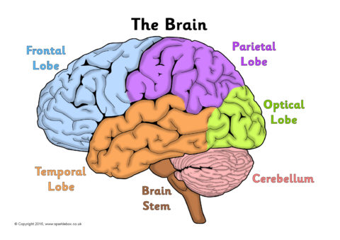
Positions and Functions of the Four Brain Lobes - MD-Health.com The brain is divided into four sections, known as lobes (as shown in the image). The frontal lobe, occipital lobe, parietal lobe, and temporal lobe have different locations and functions that support the responses and actions of the human body. Let's start by identifying where each lobe is positioned in the brain. Position of the Lobes Lobes of the brain: Structure and function | Kenhub The brain, along with the spinal cord, is the main organ of the central nervous system. It is the most complex organ of the body, with many layers and components that play their roles in almost every function performed by the body. The brain is composed of the cerebrum, cerebellum and brainstem. Brain - Human Brain Diagrams and Detailed Information The brainstem is made of three regions: the medulla oblongata, the pons, and the midbrain. A net-like structure of mixed gray and white matter known as the reticular formation is found in all three regions of the brainstem. The reticular formation controls muscle tone in the body and acts as the switch between consciousness and sleep in the brain. Label Parts of the Brain Quiz - PurposeGames.com Games by same creator. Label Parts of the Brain 17p Image Quiz. Label a Kidney 17p Image Quiz. Label the Brain and Spinal Cord 10p Image Quiz. Label a Nephron 16p Image Quiz. Label the Brain Parts 9p Image Quiz. HOTUS Midterm Map (Cities and Battles) Image Quiz.
Labeled Brain Model Diagram | Science Trends The cerebrum is the largest and most complex portion of the human brain. The cerebrum's function is to control our actions and thoughts, either conscious or unconscious, and responses to stimuli. The cerebrum itself is typically divided into four different lobes: the temporal lobe, the parietal lobe, the occipital lobe, and the frontal lobe.
Parts of the Brain: Structures, Anatomy and Functions The brain is a 3-pound organ that contains more than 100 billion neurons and many specialized areas. There are 3 main parts of the brain include the cerebrum, cerebellum, and brain stem.The Cerebrum can also be divided into 4 lobes: frontal lobes, parietal lobes, temporal lobes, and occipital lobes.The brain stem consists of three major parts: Midbrain, Pons, and Medulla oblongata.
Anatomy of the Brain: Structures and Their Function - ThoughtCo The anatomy of the brain is complex due its intricate structure and function. This amazing organ acts as a control center by receiving, interpreting, and directing sensory information throughout the body. The brain and spinal cord are the two main structures of the central nervous system. There are three major divisions of the brain.
Anatomy of the Brain | Simply Psychology The cerebrum is divided into two hemispheres, the left and right, and contains the lobes of the brain (frontal, temporal, parietal, and occipital lobes). The cerebrum produces higher functioning roles such as thinking, learning, memory, language, emotion, movement, and perception.
Label the Brain Anatomy Diagram Flashcards | Quizlet Start studying Label the Brain Anatomy Diagram. Learn vocabulary, terms, and more with flashcards, games, and other study tools.
2,783 Labeled brain anatomy Images, Stock Photos & Vectors - Shutterstock 2,783 labeled brain anatomy stock photos, vectors, and illustrations are available royalty-free. See labeled brain anatomy stock video clips Image type Orientation People Artists Sort by Popular Healthcare and Medical Anatomy Icons and Graphics human brain brain organ medicine cerebral cortex cerebellum human body Next of 28
Brain: Anatomy, Pictures, Functions, and Conditions The brainstem is an area located at the base of the brain that contains structures vital for involuntary functions such as the heartbeat and breathing. The brain stem is comprised of the midbrain, pons, and medulla. 3 Midbrain The midbrain is often considered the smallest region of the brain.
Nervous System - Label the Brain Nervous System - Label the Brain Nervous System - Brain Name: Choose the correct names for the parts of the brain. ( 1) (2) (3) (4) (5) (6) (7) (8) ( 9) This brain part controls thinking. (10) This brain part controls balance, movement, and coordination. (11) This brain part controls involuntary actions such as breathing, heartbeats, and digestion.
Amazon.com: XINDAM 3D Human Brain with Labels Anatomical Model ... This item: XINDAM 3D Human Brain with Labels Anatomical Model Paperweight (Laser Etched) in Crystal Glass Ball Science Gift (Included LED Base) $66.99 Brain 11 Ounce Ceramic Coffee Mug (WC462M) $16.98 Anatomic Brain Specimen Coasters (Set of 10) - Neuroscience Gifts, Gifts for Medical Student Gifts Brain Decor Human Anatomy Gifts
Amazon.com: brain model labeled VEVOR Human Brain Model Anatomy 4-Part Model of Brain w/Labels & Display Base Color-Coded Life Size Human Brain Anatomical Model Brain Teaching Human Brain for Science Classroom Study Display Model. 3.4 out of 5 stars 3. $159.19 $ 159. 19. Get it Wed, Mar 30 - Mon, Apr 4. FREE Shipping.
Label The Brain Worksheets & Teaching Resources | TpT The Primary Brain. 89. $2.50. Digital Download. ZIP (19.68 MB) These table caddy labels measure 8" by 3" in size and are completely EDITABLE! They can be used as classroom labels for a table caddy, table numbers, name tags, or anything else you need this size label for.
3D Brain This interactive brain model is powered by the Wellcome Trust and developed by Matt Wimsatt and Jack Simpson; reviewed by John Morrison, Patrick Hof, and Edward Lein. Structure descriptions were written by Levi Gadye and Alexis Wnuk and Jane Roskams .
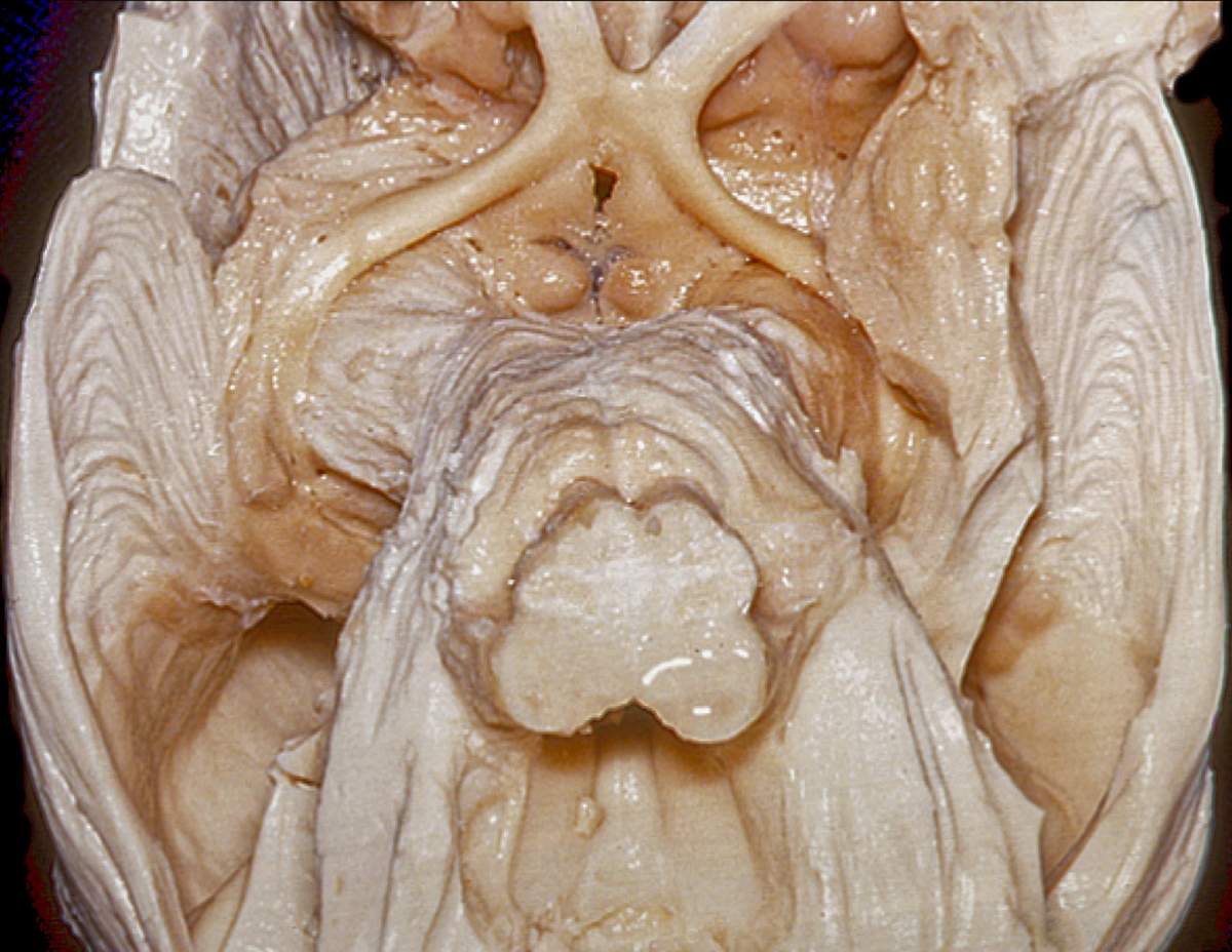
Inferior View of Visual Pathway | Neuroanatomy | The Neurosurgical Atlas, by Aaron Cohen-Gadol, M.D.
Labeled Diagrams of the Human Brain You'll Want to Copy Now Labeled Diagrams of the Human Brain Central Core The central core consists of the thalamus, pons, cerebellum, reticular formation and medulla. These five regions are the central areas that regulate breathing, pulse, arousal, balance, sleep and early stages of processing sensory information.






Post a Comment for "43 the brain with labels"